Category Archives: featured

Current Biology – Chitin in cartilaginous fishes

Our new work in collaboration with Chris Amemiya’s lab at UC Merced demonstrates the presence of Chitin in an unexpected place – the electro sensory organs of cartilaginous fishes.

Molly Phillips, W. Joyce Tang, Matthew Robinson, Daniel Ocampo Daza, Khan Hassan, Valerie Leppert, Linda S. Hirst, Chris T. Amemiya,
“Evidence of chitin in the ampullae of Lorenzini of chondrichthyan fishes”, Current Biology, Volume 30, Issue 20, 2020, Pages R1254-R1255,
https://doi.org/10.1016/j.cub.2020.08.014.

Read a news article here

https://news.ucmerced.edu/news/2020/hard-shells-and-electrosensory-gels-lab-makes-surprising-discovery

Abstract,We previously reported that the polysaccharide chitin, a key component of arthropod exoskeletons and fungal cell walls, is endogenously produced by fishes and amphibians in spite of the widely held view that it was not synthesized by vertebrates [1]. Genes encoding chitin synthase enzymes were found in the genomes of a number of fishes and amphibians and shown to be correspondingly expressed at the sites where chitin was localized [1,2]. In this report, we present evidence suggesting that chitin is prevalent within the specialized electrosensory organs of cartilaginous fishes (Chondrichthyes). These organs, the Ampullae of Lorenzini (AoL), are widely distributed and comprise a series of gel-filled canals emanating from pores in the skin ( Figure 1A). The canals extend into bulbous structures called alveoli that contain sensory cells capable of detecting subtle changes in electric fields ( Figure 1B) [3,4]. The findings described here extend the number of vertebrate taxa where endogenous chitin production has been detected and raise questions regarding chitin’s potential function in chondrichthyan fishes and other aquatic vertebrates.

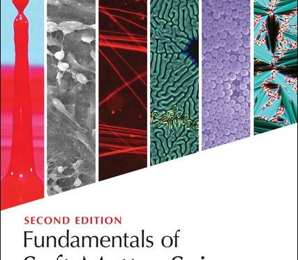
FUndamentals of Soft matter science 2nd Ed – out now!

Summary

This revised edition continues to provide the most approachable introduction to the structure, characteristics, and everyday applications of soft matter. It begins with a substantially revised overview of the underlying physics and chemistry common to soft materials. Subsequent chapters comprehensively address the different classes of soft materials, from liquid crystals to surfactants, polymers, colloids, and biomaterials, with vivid, full-color illustrations throughout. There are new worked examples throughout, new problems, some deeper mathematical treatment, and new sections on key topics such as diffusion, active matter, liquid crystal defects, surfactant phases and more.

• Introduces the science of soft materials, experimental methods used in their study, and wide-ranging applications in everyday life.

• Provides brand new worked examples throughout, in addition to expanded chapter problem sets and an updated glossary.

• Includes expanded mathematical content and substantially revised introductory chapters. 

This book will provide a comprehensive introductory resource to both undergraduate and graduate students discovering soft materials for the first time and is aimed at students with an introductory college background in physics, chemistry or materials science. 

Nanofoams – Nature communications

Nanoparticle-based hollow microstructures formed by two-stage nematic nucleation and phase separation

https://news.ucmerced.edu//news/2019/grad-student-discovers-patents-process-create-%E2%80%9Cnanofoams%E2%80%9D-liquid-crystals

Sheida T. Riahinasab, Amir Keshavarz, Charles N. Melton, Ahmed Elbaradei, Gabrielle I. Warren, Robin L. B. Selinger, Benjamin J. Stokes & Linda S. Hirst 

Nature Communications volume 10, Article number: 894 (2019)

read the paper here

Rapid bulk assembly of nanoparticles into microstructures is challenging, but highly desirable for applications in controlled release, catalysis, and sensing. We report a method to form hollow microstructures via a two-stage nematic nucleation process, generating size-tunable closed-cell foams, spherical shells, and tubular networks composed of closely packed nanoparticles. Mesogen-modified nanoparticles are dispersed in liquid crystal above the nematic-isotropic transition temperature (TNI). On cooling through TNI, nanoparticles first segregate into shrinking isotropic domains where they locally depress the transition temperature. On further cooling, nematic domains nucleate inside the nanoparticle-rich isotropic domains, driving formation of hollow nanoparticle assemblies. Structural differentiation is controlled by nanoparticle density and cooling rate. Cahn-Hilliard simulations of phase separation in liquid crystal demonstrate qualitatively that partitioning of nanoparticles into isolated domains is strongly affected by cooling rate, supporting experimental observations that cooling rate controls aggregate size. Microscopy suggests the number and size of internal voids is controlled by second-stage nucleation.

Using molecular motors to extract lipid tubules

A Simple Experimental Model to Investigate Force Range for Membrane Nanotube Formation

Chai Lor1, Joseph D. Lopes2, Michelle K. Mattson-Hoss3, Jing Xu2* and Linda S. Hirst2*
  • 1Biological Engineering and Small-scale Technologies Graduate Program, School of Engineering, University of California Merced, Merced, CA, USA
  • 2Physics Department, School of Natural Science, University of California Merced, Merced, CA, USA
  • 3Developmental and Cell Biology, School of Biological Sciences, University of California Irvine, Irvine, CA, USA

fmats-03-00006-g001The presence of membrane tubules in living cells is essential to many biological processes. In cells, one mechanism to form nanosized lipid tubules is via molecular motor induced bilayer extraction. In this paper, we describe a simple experimental model to investigate the forces required for lipid tube formation using kinesin motors anchored to 1,2-dioleoyl-sn-glycero-3-phosphocholine (DOPC) vesicles. Previous related studies have used molecular motors actively pulling on the membrane to extract a nanotube. Here, we invert the system geometry; molecular motors are used as static anchors linking DOPC vesicles to a two-dimensional microtubule network and an external flow is introduced to generate nanotubes facilitated by the drag force. We found that a drag force of ≈7 pN was sufficient for tubule extraction for vesicles ranging from 1 to 2 μm in radius. By our method, we found that the force generated by a single molecular motor was sufficient for membrane tubule extraction from a spherical lipid vesicle.

Congratulations Dr Lor!

Dr Chai Lor successfully defended his PhD thesis in the BEST (Bioengineering and small scale technologies) program on October 26th. Chai was a bioengineering undergraduate at UC Merced and a member of the first graduating class.

“Phase Behavior and Nanotube Formation in Lipid Membranes”

Biological cells are protected by a complex barrier called the lipid membrane. The lipid membrane is a soft material structure consisting of many lipid molecules held together by hydrophobic forces in an aqueous solution. Two simple experimental models were employed to investigate the role of specific lipid molecules in biological membranes. The first model investigated the phase behavior of the binary lipid mixture, 1-dipalmitoyl-2-docosahexaenoyl-sn-glycero-3-phosphoethanolamine (DHA-PE) and 1,2-dipalmitoyl-sn-glycero-3-phosphocholine (DPPC), using small-angle x-ray scattering (SAXS) and wide-angle x-ray scattering (WAXS). Our results shows that DHA-PE induces phase separation into a DHA rich liquid crystalline (Lα) phase and a DHA poor gel (Lβ’) phase at overall DHA-PE concentrations as low as 0.1mol%. In addition, we find that the structure of the Lβ’ phase, from which the DHA-PE molecules are largely excluded, is modified in the phase-separated state at low DHA-PE concentrations, with a decrease in bilayer thickness of 1.34nm for 0.1mol% at room temperature compared to pure DPPC bilayers. The second model investigated the formation of lipid nanotubes using an anchor system consisting of lipids, kinesin molecular motors, and microtubules in a flow cell. Lipid tubulation was conducted on two different membranes, 1,2-dioleoyl-sn-glycero-3-phosphocholine (DOPC) and DPPC. Lipid nanotubes were pulled from anchored giant unilamellar vesicles (GUVs) by drag force generated from the flow inside the channel. The results showed that DPPC membranes cannot generate lipid nanotubes while DOPC can, which was expected. We find that a drag force of approximately ≈7.9 pN is sufficient for tubule extraction and that it only requires 1-2 kinesin motor proteins for anchoring the GUV.

“Low concentrations of docosahexanoic acid significantly modify membrane structure and phase behavior”
C. Lor and L.S. Hirst, MEMBRANES, 5(4), 857-874 Link (2015)


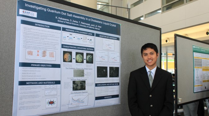
Kyle Kabasaras, CAMP summer student, presents research at UC Merced Symposium

This summer Kyle Kabasaras presented his original research project at the UC Merced undergrad research symposium.

Investigating quantum dot assembly in a cholesteric liquid crystal

An ongoing goal in condensed matter physics is directly controlling the self-assembly of quantum dots (QDs) into specific structures while maintaining their original electronic and optical properties. One method of controlling the self-assembly of QDs is to disperse them within a liquid crystal (LC) medium and apply a variety of thermal stimulations. Recently, our lab developed a method of creating spherical, vesicle-shaped QDs within a nematic LCVesicle formation depends on the QD concentration in the LC as well as the LC’s intermolecular dispersion forces and thermal properties. In this project, we investigate the dispersion of CdSe/ZnS (core/shell) QDs in a cholesteric LC (CLC) medium and predict the QD aggregations to cluster near the LC defects. By varying parameters such as QD concentration and temperature, we exploit the CLC’s sensitive optical and thermal propertiesTo observe these effects, we apply spectrophotometry, polarized optical microscopy, and fluorescence microscopy. These techniques highlight the aggregation of QDs within the host CLC and identify how LC phase transitions determine where QDaggregates form. This work illustrates the possibility of new LC-based QD devices, and we will continue by exploring the lasing potential of our sample.

camplogo1
CAMP is a National Science Foundation funded project to promote undergraduate research participation.